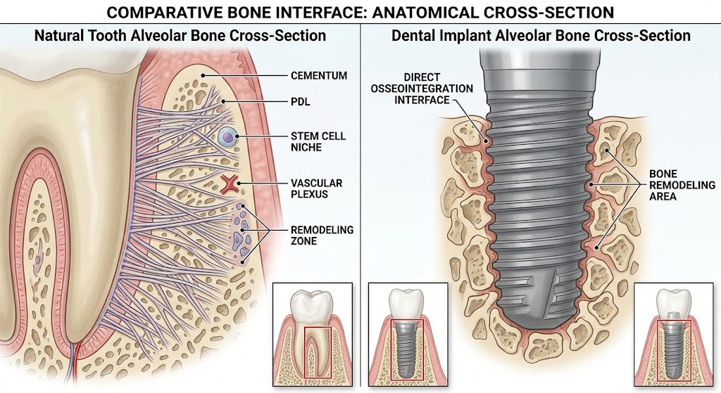
Imagine two almost identical smiles. One contains a natural tooth; the other, an implant with a crown that perfectly mimics it. The patient may not perceive any difference, but biologically, the difference is substantial. An implant can replace a missing tooth extremely successfully in both functional and esthetic terms, but it cannot completely reconstruct the tooth’s natural supporting apparatus. This is where the explanation begins as to why the tissues around an implant may be more vulnerable to the long-term presence of bacterial biofilm. A natural tooth is not simply a piece of hard tissue embedded in bone. Between the root and the alveolar bone lies the periodontal ligament, a complex, highly vascularized and innervated tissue rich in cells with significant regenerative potential. Its collagen fibers insert into the root cementum at one end and into the alveolar bone at the other, creating a true biologic attachment system. An implant functions in an entirely different way. In successful osseointegration, its surface establishes direct contact with bone, but no periodontal ligament forms around it. Osseointegration is one of the greatest achievements of modern implant dentistry, but at the same time, it means that an implant lacks one of the important anatomic structures present around a natural tooth. The difference becomes even more interesting when we examine the soft tissue. Around a natural tooth, there are complex supracrestal bundles of collagen fibers, including fibers that establish a firm attachment to the cementum. The periodontal ligament is also richly vascularized and contains fibroblasts, cementoblasts, osteoblasts, osteoclasts, and other cellular elements involved in remodeling and regeneration. No such apparatus exists around an implant. Instead, between the apical end of the junctional epithelium and the crestal bone lies a zone of connective tissue whose inner portion may be dense, relatively poorly vascularized, and organized in a manner resembling scar tissue. In many experimental descriptions, the collagen fibers are predominantly oriented parallel or circumferentially to the implant, although the literature also describes other patterns of fiber orientation. Around a natural tooth, fibers that attach to the cementum create a complex connective-tissue architecture, whereas around an implant there is no cementum into which the same periodontal fiber system could insert. In other words, the soft tissue around an implant forms a biologic barrier, but it is not a complete replica of the periodontal apparatus of a natural tooth. Nevertheless, an implant is not completely unprotected. Oral epithelium, sulcular epithelium, and junctional epithelium are present around it, and the junctional epithelium can establish an attachment to the implant surface through structures that ultrastructurally resemble the basal lamina and hemidesmosomes of a natural tooth. It is therefore incorrect to say that the body treats an implant as an unprotected piece of metal. The body creates a sophisticated mucosal barrier around it. The problem is that this barrier functions in a different anatomic environment. When biofilm accumulates on the implant surface, bacterial products come into contact with tissues that initiate an innate immune response. In an early peri-implant mucositis lesion, the numbers of neutrophils, macrophages, and T lymphocytes increase, vascular permeability increases, and loss of collagen structures begins in the subepithelial connective tissue. If the biofilm is not controlled, the inflammatory infiltrate becomes denser and extends laterally and apically. B lymphocytes and plasma cells appear, while inflammatory mediators promote further fibroblast damage, collagen loss, and the development of conditions that can activate osteoclastic mechanisms. Between the inflammatory process and the crestal bone around the implant, only a relatively thin layer of noninfiltrated connective tissue may remain. This means that persistent inflammation around an implant should not be viewed as a harmless version of gingivitis around a natural tooth. The peri-implant mucosa may appear relatively quiet for a long time while a highly active immune process is taking place at the microscopic level. Another important point is that the junctional epithelium itself is not a passive protective structure. Around a natural tooth, it also forms part of the immune boundary with the oral microbiota, allowing an exchange of bacterial products and components of the host defense system. A similar epithelial component exists around an implant, but beneath it there is no periodontal ligament with its characteristic vascular, cellular, and fibrous apparatus. Therefore, the statement that an implant has “weaker defense” should be understood precisely: an implant is not immunologically helpless, but its peri-implant tissues have a different and, in some respects, less robust anatomic organization than the tissues surrounding a natural tooth. This is why good implant dentistry does not end when the crown is placed and the patient leaves the office. True long-term success begins at that point. Biofilm control, hygienic access to the prosthetic restoration, monitoring of the peri-implant mucosa, and regular follow-up visits are not additions to treatment; they are part of implant biology. An implant may function flawlessly for decades, but we should not expect it to possess all the protective mechanisms of a tooth that evolution developed as an integral part of the periodontal complex of the natural tooth. The most dangerous misconception, therefore, is not that an implant is an artificial tooth. The most dangerous misconception is treating it as though it were biologically identical to a natural tooth.
Natural Tooth vs Implant: Why Is the Biological Defense Weaker?
Leave a Reply