- What Is the Supracrestal Tissue Attachment?
- How Much Vertical Space Is Required?
- What Happens When Space Is Insufficient?
- Clinical Implications for Implant Placement
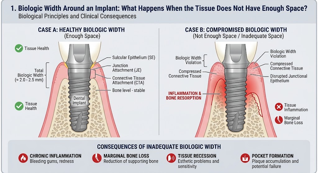
An implant may be perfectly osseointegrated, the prosthetic restoration may look excellent, and yet a process may still begin around its neck that alters the level of the marginal bone. The reason is simple: the soft tissue around an implant must form its own protective biologic attachment, and it needs space to do so. If there is not enough space, the body cannot simply bypass this requirement. It will attempt to create that space, and the crestal bone often pays the price. Biologic width represents the vertical zone between the most coronal portion of the epithelial attachment and the alveolar bone. Around an implant, this zone consists of approximately one millimeter of epithelial attachment and approximately one millimeter of supracrestal connective tissue, with the peri-implant sulcus located above them. When the entire soft-tissue complex is considered, approximately three millimeters of supracrestal space is required for the tissues to organize into a stable barrier. This is not an arbitrary measurement, but a consequence of the way the epithelium and connective tissue establish a boundary between the oral environment and the deeper peri-implant structures. The problem begins when the implant or its components are positioned in such a way that insufficient space remains for this barrier. The body cannot compress the required structures simply because we have placed the implant at a certain depth. Instead, remodeling of the crestal bone may begin. The bone shifts apically until sufficient space is created for the epithelial and connective-tissue components to assume a more stable position. This is why early marginal bone loss around an implant does not automatically indicate peri-implantitis. Under certain circumstances, it may represent a biologic adaptation through which the body reestablishes the required supracrestal tissue dimension. This is precisely where an important diagnostic pitfall arises: radiographic bone loss is not synonymous with inflammatory disease. Peri-implantitis involves much more than the simple fact that the bone level is lower than it was previously. Inflammation of the peri-implant mucosa, probing findings, the course of the changes, and progression of supporting bone loss must all be analyzed. Biologic width around an implant is also not completely identical to that around a natural tooth. Around a natural tooth, collagen fibers of the periodontal complex can insert into the cementum, whereas an implant has neither cementum nor a periodontal ligament. The connective-tissue fibers adjacent to an implant are organized differently and do not form the same insertion system that exists around the root of a natural tooth. For this reason, soft-tissue contact with an implant represents a distinct biologic structure. The junctional epithelium can establish an attachment to the implant surface through the basal lamina and hemidesmosomes, but this is not the same as fibrous attachment to the cementum of a natural tooth. The stability of the supracrestal soft tissue is therefore extremely important for protecting the deeper peri-implant region. Experimental studies have shown that adequate supracrestal space is necessary for stable organization of this complex and that, when this space is insufficient, crestal bone resorption may be expected. In other words, the bone may recede until the soft tissue acquires the space it requires. This leads to one of the most important messages in implant dentistry: the body will remodel the marginal bone rather than abandon the formation of a physiologic soft-tissue barrier. However, the problem does not end there. When the bone recedes, a portion of the implant surface that was previously surrounded by bone may become supracrestal. If that surface is rough and difficult to access for oral hygiene, this may facilitate biofilm retention. Thus, adaptive remodeling that is not itself peri-implantitis may create an anatomic situation that is more favorable for future inflammation. This is precisely why the position of the implant–abutment connection, the height of the peri-implant soft tissue, the design of the implant neck, and the vertical position of the platform are not insignificant technical details. They determine the space within which the body will attempt to build its protective soft-tissue complex. Histologic findings demonstrate how pronounced this adaptation may be: when the initial biologic width is insufficient, the crestal bone may resorb, sometimes producing a characteristic funnel-shaped change around the implant neck. It is also important to recognize that, once formed, the soft-tissue barrier is not a structure that can be manipulated indefinitely without consequences. Removing and reconnecting the abutment may disrupt the existing attachment and require the tissues to reorganize. For this reason, every intervention in this region should have a clear rationale rather than being performed as a routine habit. Additional attention has been drawn to the concept of platform switching, in which the implant–abutment connection is shifted horizontally toward the center of the implant platform. The idea is to move the inflammatory zone associated with the connection away from the marginal bone while simultaneously providing a different spatial organization of the soft tissues. Nevertheless, no design can override fundamental biology if the implant is placed in an unfavorable three-dimensional position. Implant placement depth is therefore not merely a prosthetic or esthetic issue. A component placed too deeply may alter the relationship between the soft tissue and bone, whereas a position that is too superficial may create entirely different esthetic and functional problems. In the esthetic zone, the consequences are particularly important because changes in the marginal bone may alter the mucosal contour and papilla position. A sound implant treatment plan therefore does not end with the question of where there is sufficient bone to place the implant. It must also address where the bone will be located after healing, where the connective tissue will terminate, where the epithelial attachment will be positioned, and at what level the prosthetic connection will lie. When you examine the marginal bone around an implant on a radiograph, you are actually looking at one of the boundaries of an active biologic system that is constantly attempting to establish equilibrium among bone, soft tissue, the implant surface, and the oral environment. If you provide this system with adequate space, it has a chance to stabilize. If you deprive it of that space, biology will not accept our plan simply because the implant has been technically well placed. The body will attempt to create the space it requires, and very often it will create that space precisely where we least want to lose it—at the expense of the crestal peri-implant bone.
Leave a Reply