- What Happens Immediately After Extraction?
- How Does New Bone Form Inside the Socket?
- Why Does the Ridge Still Shrink?
- What Changes During the First Eight Weeks?
- Why This Matters for Implant Planning
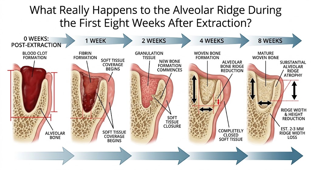
When a tooth is extracted, what remains is not simply an empty extraction socket waiting to be filled with new bone. At that moment, one of the most dynamic remodeling processes in oral surgery begins, and what is particularly important is that two processes that initially appear contradictory occur simultaneously: the body forms new bone within the socket while at the same time resorbing part of the bony walls that originally formed that socket. This is why, eight weeks after extraction, the socket may be largely filled with newly formed bone while the alveolar ridge is nevertheless narrower and lower than it was on the day of extraction. This paradox is fundamental to understanding extraction wound healing. Immediately after extraction, the socket fills with blood and a blood clot forms. The clot is not merely a plug that stops bleeding, but a temporary biologic scaffold on which the organization of future tissue begins. The fibrin network contains erythrocytes and platelets, and inflammatory cells begin to enter the wound area as early as the first day. This is not a complication, but part of normal healing. The body must clear the damaged area while simultaneously preparing the site for new vascularization and regeneration. By approximately the third day, the marginal portions of the blood clot begin to be replaced by highly vascularized connective tissue. In other words, what looked like a clot the day before begins to transform into living tissue permeated by new blood vessels. The central and apical portions of the socket still lag behind this process, demonstrating that healing is not uniform and that different parts of the same extraction wound may be in different biologic phases at the same time. By approximately the seventh day, the change becomes even more dramatic. The blood clot and granulation tissue become organized through neovascularization, undifferentiated mesenchymal cells, leukocytes, and collagen fibers growing inward from the periphery. The socket is no longer a passive cavity. It becomes an intensely active construction site. Blood vessels are particularly important because reliable bone regeneration cannot occur without angiogenesis. They not only deliver oxygen and nutrients but also create the biologic environment in which osteoprogenitor cells can differentiate into osteoblasts. At the same time, osteoclast activity also increases. This is a detail that is often overlooked: bone healing does not mean bone formation alone. Regeneration involves precisely coordinated resorption of existing structures and formation of new ones. By the second week, the marginal portion of the socket begins to acquire an epithelial covering, while the highly vascularized mesenchymal component increasingly occupies the space previously filled by the blood clot. Remnants of the periodontal ligament and the alveolar bone plate undergo degradation, creating communications with the bone marrow of the surrounding cancellous bone. It is through this vascular and cellular network that a more substantial phase of osteogenesis begins. Woven bone appears in the central portions of the socket. It forms rapidly, but it is not yet the mature lamellar bone we want to see as the final result of healing. Woven bone represents a temporary structure that the body will later remodel and replace with a more organized lamellar structure. By approximately the thirtieth day, the wound surface may already appear surprisingly calm. The marginal portion is covered by organized connective tissue and keratinized stratified squamous epithelium. Beneath the surface, however, remodeling remains highly active. The first primary osteons appear within the newly formed woven bone, marking the beginning of its maturation, while numerous osteoclasts participate in the remodeling of both newly formed and preexisting bone. In other words, one month after extraction, the bone has not finished healing; it is only beginning to undergo intensive changes in its internal architecture. At the same time, a process that may be even more important to the implant dentist than the filling of the socket itself begins to become apparent: a change in the external dimensions of the ridge. The alveolar ridge exists because the tooth exists. After the tooth is removed, part of the bony anatomy loses its original biologic and functional role, and the body begins to remodel it. Resorption occurs on both the buccal and oral aspects, but it is not symmetrical. The buccal bone plate is particularly susceptible to change, and more pronounced horizontal and vertical losses may therefore develop on this side. By approximately the fifth week, narrowing of the ridge and flattening of the buccal bone plate may already be clearly evident. This is clinically extremely important because the patient may have a completely closed wound and the impression that the area has healed, while beneath the mucosa the geometry of the ridge has already changed substantially. By the eighth week, the extraction socket may be largely filled with woven bone, but this in no way means that the anatomy has returned to its pre-extraction condition. On the contrary, very pronounced dimensional changes may occur during this early period, particularly on the buccal aspect. Experimental models show that osteoclast activity during this period may lead to substantial resorption of the crestal portions of the buccal and lingual walls, with more pronounced changes on the buccal side. Even more strikingly, after eight weeks the sockets may be filled with woven bone while vertical buccal bone loss simultaneously amounts to several millimeters. The body heals the wound very successfully, but it has no biologic reason to preserve the identical external architecture of the bone that existed while the tooth was present. This is why atraumatic extraction makes sense, but even a perfectly performed extraction cannot, by itself, completely prevent physiologic remodeling. Likewise, immediate implant placement does not provide magical protection against resorption of the buccal bone plate. After extraction, biology continues its remodeling program regardless of our prosthetic plans. The most important message of the first eight weeks, therefore, is not how quickly the space left by the extracted tooth fills with bone, but what kind of ridge will remain once the most intensive early phase of remodeling has ended. In just a few weeks, the blood clot becomes a vascularized matrix, the matrix becomes woven bone, maturation begins, the surface becomes epithelialized, and at the same time the external contour of the ridge decreases. This is why, in contemporary implant dentistry, tooth extraction should not be viewed as the end of one treatment. It is the beginning of a biologic process that will determine the anatomy of the future implant site.