It can, but not in the oversimplified way this is often presented. The mere fact that a tooth was once extracted with difficulty or traumatically does not mean that the patient will necessarily develop peri-implantitis years later. However, the extraction technique can alter the anatomy of the alveolar ridge, and that anatomy may later determine how appropriately the implant can be positioned, how much bone support it will have, and how stable the peri-implant tissues will be. In other words, an error during extraction can leave an anatomic consequence that becomes clinically important only later. When a tooth is extracted, the socket does not remain unchanged. Physiologic remodeling begins, during which the ridge gradually narrows and loses some of its height. The buccal bone plate is particularly susceptible, and changes may be more pronounced on this aspect. If part of the alveolar wall is additionally fractured or lost during extraction, the initial anatomy becomes even less favorable. The body will heal such a defect as well, but it will not necessarily restore the ridge to its original width and shape. For this reason, atraumatic extraction is not simply a more elegant surgical technique. Its essence lies in preserving what will later form the foundation for implant treatment. Careful extraction can better preserve the alveolar ridge and thereby reduce the additional loss of bone architecture that accompanies natural post-extraction remodeling. This does not mean, however, that resorption can be completely prevented. Even when the socket walls are preserved, the ridge changes after extraction. But there is an enormous difference between controlled physiologic remodeling and a situation in which important buccal or interproximal bone is lost during extraction. The problem often becomes apparent only when implant placement is being planned. If the ridge is too narrow, the surgeon must choose between augmentation, a compromised implant position, or a combination of several procedures. This is precisely where an earlier extraction can indirectly affect future risk. An implant placed in an unfavorable three-dimensional position is more difficult to clean, the prosthetic superstructure may become less favorable for oral hygiene, and part of the rough implant surface may end up closer to the soft tissue or become exposed after remodeling. Such a situation does not cause peri-implantitis by itself, because biofilm remains the central etiologic driver of the inflammatory disease, but it can create a local environment in which plaque accumulates more easily and is more difficult to remove. It is therefore important to distinguish between a cause and factors that increase the vulnerability of a site. A traumatic extraction is not a bacterial infection around a future implant, but it may contribute to an anatomy that reduces the margin of safety. Another problem arises when bone augmentation is required because of ridge loss. Regenerative procedures can reconstruct volume very successfully, but the result is not the same as preserved natural anatomy. The amount of newly formed bone, its organization, the relationship between the graft and native bone, and the stability of the contour can vary. This means that sometimes the best implant site is one that never had to be reconstructed because as much of the existing bone as possible was preserved during extraction. This is particularly important in the esthetic zone, where a few millimeters of lost buccal support can alter the soft-tissue contour, the position of the gingival margin, and the emergence profile of the future crown. But the esthetic problem is not the only concern. Thin or insufficient buccal bone may be more susceptible to further remodeling over time, and if recession occurs, the rough implant surface may become more accessible to oral biofilm. What began as an anatomic compromise can then become a factor that complicates maintenance of peri-implant health. It is important, however, not to go to the opposite extreme. There is no scientific justification for attributing peri-implantitis that develops years later in every patient to an extraction performed long ago. Peri-implantitis is a multifactorial problem in which biofilm, a history of periodontitis, oral hygiene, smoking, prosthetic design, implant position, and local conditions are much more direct factors. The way a tooth was extracted should be viewed as a possible first step in a chain of events, not as an independent cause of disease. The most important question, therefore, is not whether the tooth was extracted quickly or slowly, but what remained of the ridge after extraction. If the bony walls are preserved, the implant clinician later has more room to achieve an appropriate three-dimensional implant position, a more stable soft-tissue contour, and a prosthetic design that is easier to maintain. If the anatomy is severely compromised, every subsequent step becomes more demanding. This is precisely why the moment of tooth extraction may be far more important for the future implant than the patient might assume. The implant may not be placed for months or years, but the bone encountered at that time began taking on its future shape on the very day the tooth was extracted. In implant dentistry, therefore, a well-executed extraction is not merely the end of treatment for one tooth. Very often, it is the first surgical step in protecting the site where an implant may be placed years later.
Author: stomatolog77
-
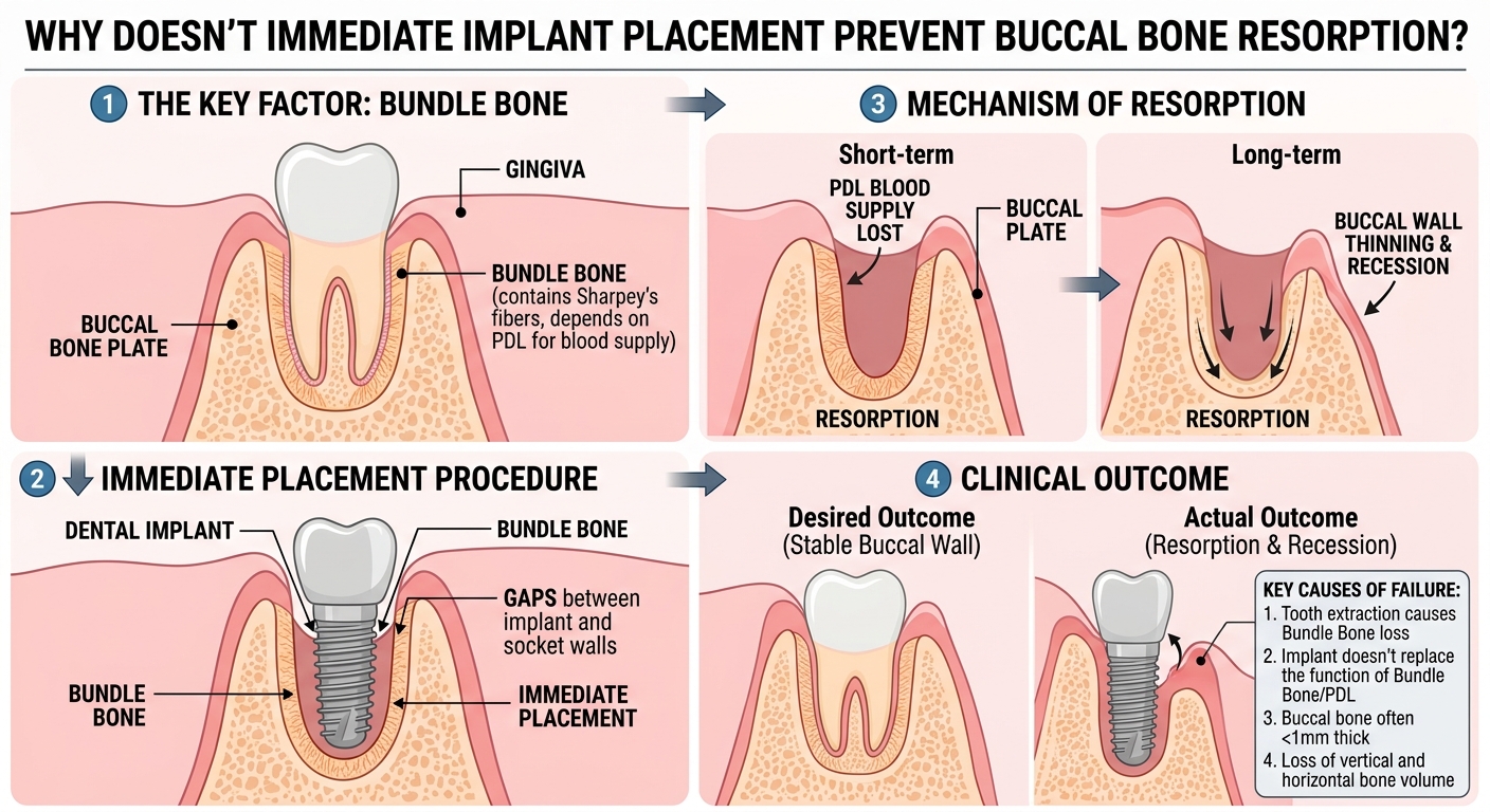
Why Doesn’t Immediate Implant Placement Prevent Buccal Bone Resorption?
- Why the Buccal Plate Resorbs
- What Immediate Implant Placement Can and Cannot Do
- Why Primary Stability Is Not Enough
- Clinical Implications for Implant Positioning
The greatest misconception about immediate implant placement is the idea that an implant, simply because it is placed immediately after extraction, will “hold” the socket and prevent it from shrinking. It will not. An implant may perfectly occupy the space previously occupied by the root, achieve excellent primary stability, and later become fully osseointegrated, while the buccal bone plate still undergoes substantial resorption. The reason is simple but biologically very important: an implant does not stop the remodeling process that begins the moment a tooth is extracted. Extraction does not merely remove the crown and root; it changes the entire biologic environment of the alveolar process. The tissues that were organized around the natural tooth begin to heal, the bony walls undergo remodeling, and the dimensions of the ridge begin to change regardless of whether an implant is placed immediately into the socket or the socket is left to heal. This is precisely why immediate implant placement must not be equated with preservation of the alveolar ridge. Experimental observations have shown that after extraction, resorption occurs on both the buccal and oral aspects of the socket, but is more pronounced buccally. This means that remodeling is not symmetrical. The buccal aspect may lose more height and width, directly altering the anatomy of the future peri-implant tissues. What is particularly interesting is that this process may continue even when an implant is placed immediately into a fresh extraction socket. In other words, a titanium implant is not a biologic replacement for a natural root in terms of preserving the surrounding bone anatomy. It can replace the function of a future support for a prosthetic restoration, but it cannot reverse the response of the alveolar ridge to tooth loss. Experimental models have shown that immediate placement of a screw-shaped titanium implant could not compensate for resorption of the buccal cortical plate, and substantial vertical loss of buccal bone was recorded over a period of several months. This is a very important message because it challenges the intuitive assumption that the presence of an implant will mechanically prevent collapse of the bony wall. Bone does not behave like a construction material that remains in place simply because a metal structure is positioned next to it. It is living tissue that continuously responds to changes in its local environment. After extraction, osteoclastic activity increases and the crestal portions of the socket undergo remodeling. An implant placed in the socket does not interrupt this biologic program. Consequently, the implant may remain stable while the contour of the ridge around it changes at the same time. This fact is particularly important in the esthetic zone. If the buccal bone loses volume, the consequence is not merely a radiographically lower bone level. Support for the peri-implant mucosa may change, the vestibular contour may become flatter, and the esthetic outcome may become less predictable. Therefore, the success of immediate implant placement should not be assessed solely by asking whether the implant has osseointegrated. An implant may survive while the surrounding anatomy changes in a way that compromises the esthetic outcome. Another important point is that post-extraction resorption occurs very early. Ridge narrowing and flattening of the buccal bone plate may already be observed during the first few weeks. After several weeks, the extraction socket may be filled with newly formed woven bone, while at the same time the external contour of the ridge is no longer the same as it was on the day of extraction. This is a paradox that every clinician involved in implant dentistry must understand: new bone may form within the socket while the preexisting crestal bone is simultaneously resorbed externally. For this reason, osseointegration and ridge preservation are not the same process. An immediate implant may successfully integrate into newly formed bone, but this does not mean that the original buccal anatomy has been preserved. Atraumatic extraction is therefore important because preservation of the existing bony walls reduces additional surgical trauma, but even a perfectly atraumatic extraction cannot completely stop the physiologic remodeling that follows tooth loss. The same applies to implant position itself. Immediate implant placement is not simply a matter of placing an implant exactly where the root used to be. The original socket and the ideal three-dimensional position of the future implant are not necessarily the same location. If the implant is positioned solely according to the anatomy of the extraction socket, it may end up in a position that is not optimal for the future relationship among bone, soft tissue, and the prosthetic crown. Therefore, with immediate implant placement, it is important to anticipate how the ridge will look after remodeling rather than focusing only on how it appears at the time of extraction. Additional procedures aimed at preserving or regenerating the ridge may reduce dimensional changes, but they do not make the biology completely predictable. Even different grafts and materials may exhibit different patterns of resorption, integration, and healing. The key point is that there is no simple intervention that can guarantee that the buccal bone will remain completely unchanged. Immediate implant placement primarily shortens the treatment pathway and allows the implant to be placed at the time of extraction. This is a major clinical advantage, but it is a mistake to conclude that alveolar resorption has therefore been stopped at the same time. The most important message is this: an implant placed immediately after tooth extraction does not “freeze” the existing anatomy. The buccal bone continues to remodel because the body is responding to the loss of the natural tooth, not to our prosthetic plan. Therefore, successful immediate implant placement does not begin with the question, “Can I place the implant immediately?” but with a far more important one: “What will happen to the buccal wall once the natural post-extraction biology has run its course?”
-
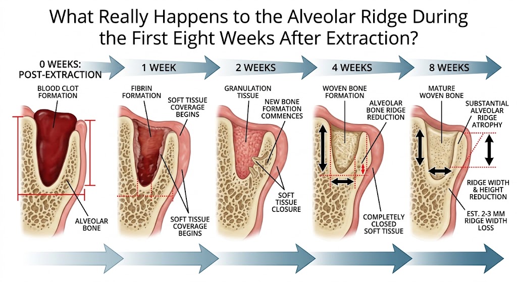
What Happens to the Alveolar Ridge After Extraction?
- What Happens Immediately After Extraction?
- How Does New Bone Form Inside the Socket?
- Why Does the Ridge Still Shrink?
- What Changes During the First Eight Weeks?
- Why This Matters for Implant Planning
When a tooth is extracted, what remains is not simply an empty extraction socket waiting to be filled with new bone. At that moment, one of the most dynamic remodeling processes in oral surgery begins, and what is particularly important is that two processes that initially appear contradictory occur simultaneously: the body forms new bone within the socket while at the same time resorbing part of the bony walls that originally formed that socket. This is why, eight weeks after extraction, the socket may be largely filled with newly formed bone while the alveolar ridge is nevertheless narrower and lower than it was on the day of extraction. This paradox is fundamental to understanding extraction wound healing. Immediately after extraction, the socket fills with blood and a blood clot forms. The clot is not merely a plug that stops bleeding, but a temporary biologic scaffold on which the organization of future tissue begins. The fibrin network contains erythrocytes and platelets, and inflammatory cells begin to enter the wound area as early as the first day. This is not a complication, but part of normal healing. The body must clear the damaged area while simultaneously preparing the site for new vascularization and regeneration. By approximately the third day, the marginal portions of the blood clot begin to be replaced by highly vascularized connective tissue. In other words, what looked like a clot the day before begins to transform into living tissue permeated by new blood vessels. The central and apical portions of the socket still lag behind this process, demonstrating that healing is not uniform and that different parts of the same extraction wound may be in different biologic phases at the same time. By approximately the seventh day, the change becomes even more dramatic. The blood clot and granulation tissue become organized through neovascularization, undifferentiated mesenchymal cells, leukocytes, and collagen fibers growing inward from the periphery. The socket is no longer a passive cavity. It becomes an intensely active construction site. Blood vessels are particularly important because reliable bone regeneration cannot occur without angiogenesis. They not only deliver oxygen and nutrients but also create the biologic environment in which osteoprogenitor cells can differentiate into osteoblasts. At the same time, osteoclast activity also increases. This is a detail that is often overlooked: bone healing does not mean bone formation alone. Regeneration involves precisely coordinated resorption of existing structures and formation of new ones. By the second week, the marginal portion of the socket begins to acquire an epithelial covering, while the highly vascularized mesenchymal component increasingly occupies the space previously filled by the blood clot. Remnants of the periodontal ligament and the alveolar bone plate undergo degradation, creating communications with the bone marrow of the surrounding cancellous bone. It is through this vascular and cellular network that a more substantial phase of osteogenesis begins. Woven bone appears in the central portions of the socket. It forms rapidly, but it is not yet the mature lamellar bone we want to see as the final result of healing. Woven bone represents a temporary structure that the body will later remodel and replace with a more organized lamellar structure. By approximately the thirtieth day, the wound surface may already appear surprisingly calm. The marginal portion is covered by organized connective tissue and keratinized stratified squamous epithelium. Beneath the surface, however, remodeling remains highly active. The first primary osteons appear within the newly formed woven bone, marking the beginning of its maturation, while numerous osteoclasts participate in the remodeling of both newly formed and preexisting bone. In other words, one month after extraction, the bone has not finished healing; it is only beginning to undergo intensive changes in its internal architecture. At the same time, a process that may be even more important to the implant dentist than the filling of the socket itself begins to become apparent: a change in the external dimensions of the ridge. The alveolar ridge exists because the tooth exists. After the tooth is removed, part of the bony anatomy loses its original biologic and functional role, and the body begins to remodel it. Resorption occurs on both the buccal and oral aspects, but it is not symmetrical. The buccal bone plate is particularly susceptible to change, and more pronounced horizontal and vertical losses may therefore develop on this side. By approximately the fifth week, narrowing of the ridge and flattening of the buccal bone plate may already be clearly evident. This is clinically extremely important because the patient may have a completely closed wound and the impression that the area has healed, while beneath the mucosa the geometry of the ridge has already changed substantially. By the eighth week, the extraction socket may be largely filled with woven bone, but this in no way means that the anatomy has returned to its pre-extraction condition. On the contrary, very pronounced dimensional changes may occur during this early period, particularly on the buccal aspect. Experimental models show that osteoclast activity during this period may lead to substantial resorption of the crestal portions of the buccal and lingual walls, with more pronounced changes on the buccal side. Even more strikingly, after eight weeks the sockets may be filled with woven bone while vertical buccal bone loss simultaneously amounts to several millimeters. The body heals the wound very successfully, but it has no biologic reason to preserve the identical external architecture of the bone that existed while the tooth was present. This is why atraumatic extraction makes sense, but even a perfectly performed extraction cannot, by itself, completely prevent physiologic remodeling. Likewise, immediate implant placement does not provide magical protection against resorption of the buccal bone plate. After extraction, biology continues its remodeling program regardless of our prosthetic plans. The most important message of the first eight weeks, therefore, is not how quickly the space left by the extracted tooth fills with bone, but what kind of ridge will remain once the most intensive early phase of remodeling has ended. In just a few weeks, the blood clot becomes a vascularized matrix, the matrix becomes woven bone, maturation begins, the surface becomes epithelialized, and at the same time the external contour of the ridge decreases. This is why, in contemporary implant dentistry, tooth extraction should not be viewed as the end of one treatment. It is the beginning of a biologic process that will determine the anatomy of the future implant site.
-
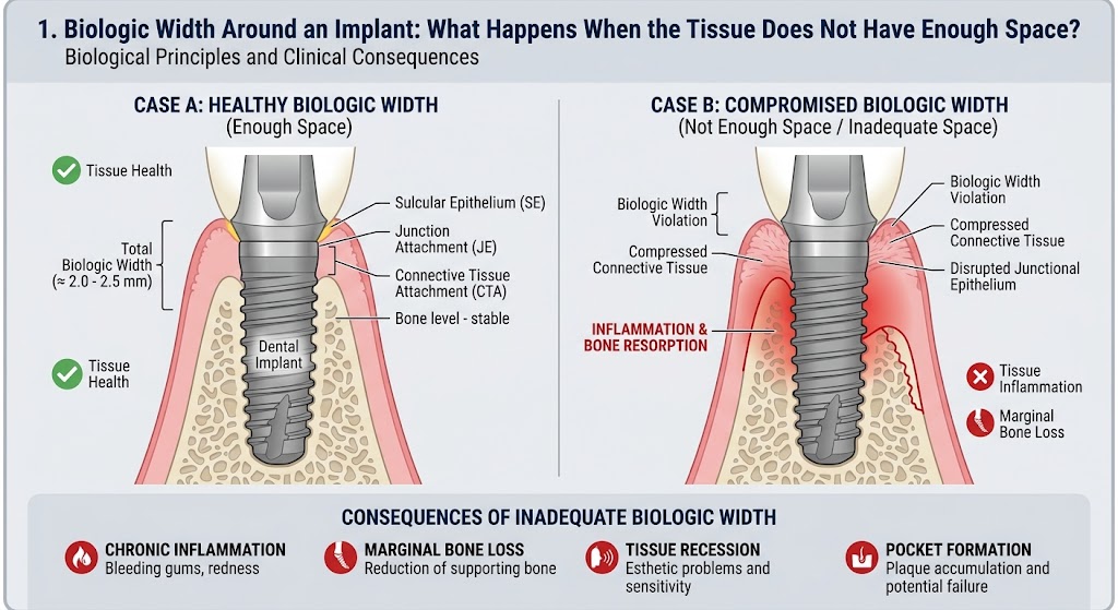
Biologic Width Around an Implant: What Happens When the Tissue Does Not Have Enough Space?
- What Is the Supracrestal Tissue Attachment?
- How Much Vertical Space Is Required?
- What Happens When Space Is Insufficient?
- Clinical Implications for Implant Placement
An implant may be perfectly osseointegrated, the prosthetic restoration may look excellent, and yet a process may still begin around its neck that alters the level of the marginal bone. The reason is simple: the soft tissue around an implant must form its own protective biologic attachment, and it needs space to do so. If there is not enough space, the body cannot simply bypass this requirement. It will attempt to create that space, and the crestal bone often pays the price. Biologic width represents the vertical zone between the most coronal portion of the epithelial attachment and the alveolar bone. Around an implant, this zone consists of approximately one millimeter of epithelial attachment and approximately one millimeter of supracrestal connective tissue, with the peri-implant sulcus located above them. When the entire soft-tissue complex is considered, approximately three millimeters of supracrestal space is required for the tissues to organize into a stable barrier. This is not an arbitrary measurement, but a consequence of the way the epithelium and connective tissue establish a boundary between the oral environment and the deeper peri-implant structures. The problem begins when the implant or its components are positioned in such a way that insufficient space remains for this barrier. The body cannot compress the required structures simply because we have placed the implant at a certain depth. Instead, remodeling of the crestal bone may begin. The bone shifts apically until sufficient space is created for the epithelial and connective-tissue components to assume a more stable position. This is why early marginal bone loss around an implant does not automatically indicate peri-implantitis. Under certain circumstances, it may represent a biologic adaptation through which the body reestablishes the required supracrestal tissue dimension. This is precisely where an important diagnostic pitfall arises: radiographic bone loss is not synonymous with inflammatory disease. Peri-implantitis involves much more than the simple fact that the bone level is lower than it was previously. Inflammation of the peri-implant mucosa, probing findings, the course of the changes, and progression of supporting bone loss must all be analyzed. Biologic width around an implant is also not completely identical to that around a natural tooth. Around a natural tooth, collagen fibers of the periodontal complex can insert into the cementum, whereas an implant has neither cementum nor a periodontal ligament. The connective-tissue fibers adjacent to an implant are organized differently and do not form the same insertion system that exists around the root of a natural tooth. For this reason, soft-tissue contact with an implant represents a distinct biologic structure. The junctional epithelium can establish an attachment to the implant surface through the basal lamina and hemidesmosomes, but this is not the same as fibrous attachment to the cementum of a natural tooth. The stability of the supracrestal soft tissue is therefore extremely important for protecting the deeper peri-implant region. Experimental studies have shown that adequate supracrestal space is necessary for stable organization of this complex and that, when this space is insufficient, crestal bone resorption may be expected. In other words, the bone may recede until the soft tissue acquires the space it requires. This leads to one of the most important messages in implant dentistry: the body will remodel the marginal bone rather than abandon the formation of a physiologic soft-tissue barrier. However, the problem does not end there. When the bone recedes, a portion of the implant surface that was previously surrounded by bone may become supracrestal. If that surface is rough and difficult to access for oral hygiene, this may facilitate biofilm retention. Thus, adaptive remodeling that is not itself peri-implantitis may create an anatomic situation that is more favorable for future inflammation. This is precisely why the position of the implant–abutment connection, the height of the peri-implant soft tissue, the design of the implant neck, and the vertical position of the platform are not insignificant technical details. They determine the space within which the body will attempt to build its protective soft-tissue complex. Histologic findings demonstrate how pronounced this adaptation may be: when the initial biologic width is insufficient, the crestal bone may resorb, sometimes producing a characteristic funnel-shaped change around the implant neck. It is also important to recognize that, once formed, the soft-tissue barrier is not a structure that can be manipulated indefinitely without consequences. Removing and reconnecting the abutment may disrupt the existing attachment and require the tissues to reorganize. For this reason, every intervention in this region should have a clear rationale rather than being performed as a routine habit. Additional attention has been drawn to the concept of platform switching, in which the implant–abutment connection is shifted horizontally toward the center of the implant platform. The idea is to move the inflammatory zone associated with the connection away from the marginal bone while simultaneously providing a different spatial organization of the soft tissues. Nevertheless, no design can override fundamental biology if the implant is placed in an unfavorable three-dimensional position. Implant placement depth is therefore not merely a prosthetic or esthetic issue. A component placed too deeply may alter the relationship between the soft tissue and bone, whereas a position that is too superficial may create entirely different esthetic and functional problems. In the esthetic zone, the consequences are particularly important because changes in the marginal bone may alter the mucosal contour and papilla position. A sound implant treatment plan therefore does not end with the question of where there is sufficient bone to place the implant. It must also address where the bone will be located after healing, where the connective tissue will terminate, where the epithelial attachment will be positioned, and at what level the prosthetic connection will lie. When you examine the marginal bone around an implant on a radiograph, you are actually looking at one of the boundaries of an active biologic system that is constantly attempting to establish equilibrium among bone, soft tissue, the implant surface, and the oral environment. If you provide this system with adequate space, it has a chance to stabilize. If you deprive it of that space, biology will not accept our plan simply because the implant has been technically well placed. The body will attempt to create the space it requires, and very often it will create that space precisely where we least want to lose it—at the expense of the crestal peri-implant bone.
-
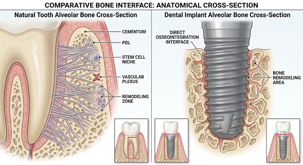
Natural Tooth vs Implant: Why Is the Biological Defense Weaker?
Imagine two almost identical smiles. One contains a natural tooth; the other, an implant with a crown that perfectly mimics it. The patient may not perceive any difference, but biologically, the difference is substantial. An implant can replace a missing tooth extremely successfully in both functional and esthetic terms, but it cannot completely reconstruct the tooth’s natural supporting apparatus. This is where the explanation begins as to why the tissues around an implant may be more vulnerable to the long-term presence of bacterial biofilm. A natural tooth is not simply a piece of hard tissue embedded in bone. Between the root and the alveolar bone lies the periodontal ligament, a complex, highly vascularized and innervated tissue rich in cells with significant regenerative potential. Its collagen fibers insert into the root cementum at one end and into the alveolar bone at the other, creating a true biologic attachment system. An implant functions in an entirely different way. In successful osseointegration, its surface establishes direct contact with bone, but no periodontal ligament forms around it. Osseointegration is one of the greatest achievements of modern implant dentistry, but at the same time, it means that an implant lacks one of the important anatomic structures present around a natural tooth. The difference becomes even more interesting when we examine the soft tissue. Around a natural tooth, there are complex supracrestal bundles of collagen fibers, including fibers that establish a firm attachment to the cementum. The periodontal ligament is also richly vascularized and contains fibroblasts, cementoblasts, osteoblasts, osteoclasts, and other cellular elements involved in remodeling and regeneration. No such apparatus exists around an implant. Instead, between the apical end of the junctional epithelium and the crestal bone lies a zone of connective tissue whose inner portion may be dense, relatively poorly vascularized, and organized in a manner resembling scar tissue. In many experimental descriptions, the collagen fibers are predominantly oriented parallel or circumferentially to the implant, although the literature also describes other patterns of fiber orientation. Around a natural tooth, fibers that attach to the cementum create a complex connective-tissue architecture, whereas around an implant there is no cementum into which the same periodontal fiber system could insert. In other words, the soft tissue around an implant forms a biologic barrier, but it is not a complete replica of the periodontal apparatus of a natural tooth. Nevertheless, an implant is not completely unprotected. Oral epithelium, sulcular epithelium, and junctional epithelium are present around it, and the junctional epithelium can establish an attachment to the implant surface through structures that ultrastructurally resemble the basal lamina and hemidesmosomes of a natural tooth. It is therefore incorrect to say that the body treats an implant as an unprotected piece of metal. The body creates a sophisticated mucosal barrier around it. The problem is that this barrier functions in a different anatomic environment. When biofilm accumulates on the implant surface, bacterial products come into contact with tissues that initiate an innate immune response. In an early peri-implant mucositis lesion, the numbers of neutrophils, macrophages, and T lymphocytes increase, vascular permeability increases, and loss of collagen structures begins in the subepithelial connective tissue. If the biofilm is not controlled, the inflammatory infiltrate becomes denser and extends laterally and apically. B lymphocytes and plasma cells appear, while inflammatory mediators promote further fibroblast damage, collagen loss, and the development of conditions that can activate osteoclastic mechanisms. Between the inflammatory process and the crestal bone around the implant, only a relatively thin layer of noninfiltrated connective tissue may remain. This means that persistent inflammation around an implant should not be viewed as a harmless version of gingivitis around a natural tooth. The peri-implant mucosa may appear relatively quiet for a long time while a highly active immune process is taking place at the microscopic level. Another important point is that the junctional epithelium itself is not a passive protective structure. Around a natural tooth, it also forms part of the immune boundary with the oral microbiota, allowing an exchange of bacterial products and components of the host defense system. A similar epithelial component exists around an implant, but beneath it there is no periodontal ligament with its characteristic vascular, cellular, and fibrous apparatus. Therefore, the statement that an implant has “weaker defense” should be understood precisely: an implant is not immunologically helpless, but its peri-implant tissues have a different and, in some respects, less robust anatomic organization than the tissues surrounding a natural tooth. This is why good implant dentistry does not end when the crown is placed and the patient leaves the office. True long-term success begins at that point. Biofilm control, hygienic access to the prosthetic restoration, monitoring of the peri-implant mucosa, and regular follow-up visits are not additions to treatment; they are part of implant biology. An implant may function flawlessly for decades, but we should not expect it to possess all the protective mechanisms of a tooth that evolution developed as an integral part of the periodontal complex of the natural tooth. The most dangerous misconception, therefore, is not that an implant is an artificial tooth. The most dangerous misconception is treating it as though it were biologically identical to a natural tooth.